Dr. Williams' Urology Resource
A Guide to Men's Health, Reproductive Medicine, and Microsurgery
Testicular Pain
Chronic testicular pain is a frustrating condition for both patients and doctors. It can be constant, can come and go, can be present in one or both testicles, and can significantly impact quality of life.
The first step in evaluating testicular pain is to identify any potentially harmful cause of the pain, such as testicular cancer, infection, or spinal injury. Rarely, a kidney stone may cause testicular pain.
A testicular ultrasound is commonly performed to evaluate for conditions including varicocele, hydrocele, spermatocele, or small testicular tumors that are difficult to feel on examination.
All too often, however, no identifiable cause of the testicular pain is found, leaving patients and providers confused and frustrated.
Intial treatments may include antibiotics, anti-inflammatory medications, narcotic pain pills, antidepressants, or anticonvulsants.
More advanced treatments may include nerve blocks, pelvic floor physical therapy, biofeedback, acupuncture, and psychotherapy.
Before presenting to a urologist, many patients have the condition for years and have been evaluated by multiple health care providers.
In the past, when intial treatments failed to offer relief, few options existed short of orchiectomy (removal of the entire testicle) which often caused physiological problems, such as low testosterone, as well as psychological distress.
Today, however, fellowship-trained microsurgeons can offer patients Microsurgical Denervation of the Spermatc Cord (MDSC). This minimally-invasive procedure allows for relief of pain and preserves testicular function.
Prior to surgery, patients are given a nerve block in the office. If pain improves with the nerve block, then patients are offered MDSC.
While no procedure is 100% effective, up to 80-90% of patients report durable improvement and relief of their testicular pain.
Microsurgical Denervation of the Spermatic Cord
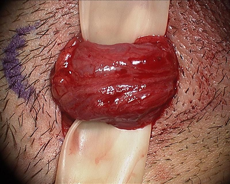
The spermatic cord is delivered into the operating field via a small, 2-3 cm incision.

Next, using the operating microscope for high-powered optical magnification and also using the microsopic doppler ultrasound, careful and meticulous dissection is carried our, and critical structures are isolated.

Finally, all remaining structures (including the nerves) are ligated. The previously-isolated critical structures--the arteries, lymphatics, and in this particular case, the vas deferens (red vessel loop)--are spared.
Watch Dr. Williams Perform an MDSC